Research Highlights
Biophysics | Other
Probing Proton Pumping: New Findings on Protein Folding in bacteriorhodopsin (bR)
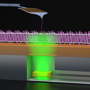
Published: February 05, 2024
PI: Thomas Perkins
Biophysics
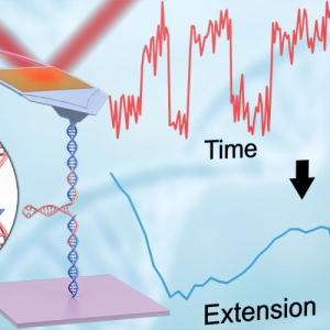
The Forces involved in Folding Proteins
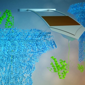
Published: March 22, 2021
PI: Thomas Perkins
Biophysics | Precision Measurement
DNA imaging, ready in five minutes
Published: July 16, 2019
PI: Thomas Perkins


Nanoscience
The Land of Enhancement: AFM Spectroscopy

Published: October 16, 2015
PI: Thomas Perkins

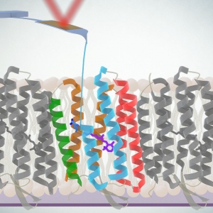
Biophysics | Nanoscience
bR Phone Home

Published: February 04, 2014
PI: Markus Raschke | PI: Thomas Perkins


 lab")
Biophysics | Nanoscience
How to Marry a Microscope

Published: April 10, 2009
PI: Thomas Perkins
Biophysics | Nanoscience
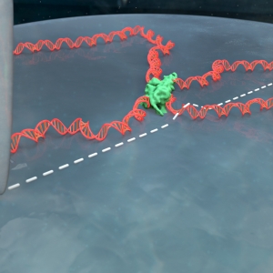
DNA: Force of Nature

Published: February 07, 2008
PI: Thomas Perkins

Biophysics | Nanoscience
Sightseeing along a DNA Strand

Published: May 01, 2005
PI: Thomas Perkins