The Kapteyn/Murnane group recently proved that you don’t need an accelerator facility to make the X-Rays for an X-Ray microscope. In fact, you can build the whole device on an optical bench — if you use a femtosecond laser to generate coherent X-Rays. The group makes coherent X-Rays by shining the laser into a glass tube filled with argon gas. The argon atoms absorb many low-energy laser photons and spit out high-energy X-Ray photons when they give up the absorbed energy. The X-Ray beam has all the desirable properties of laser light. For example, it does not spread rapidly and can be used to make holograms. (see "X-Ray Vision" in JILA: Light & Matter, Spring 2007).
JILA’s new X-Ray microscope offers the promise of high-resolution imaging of micron-scale materials and structures, entire cells, mitochondria, and possibly even DNA or the proteins in cell membranes. It will be particularly important to the fields of biology and medicine because it can image thick samples, unlike a scanning electron microscope that can only image surfaces. "In 10–20 years when the X-Ray microscope is fully developed, every hospital may have one for diagnosing different illnesses via high-resolution images of single cells," Kapteyn predicts.
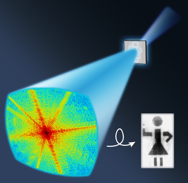
JILA’s X-Ray microscope was invented by graduate students Richard Sandberg, Daisy Raymondson, and Chan La-O-Vorakiat, research associate Ariel Paul, Fellows Margaret Murnane and Henry Kapteyn, and colleagues from UCLA, CSU, Lawrence Berkeley National Laboratory, and the Universities of California at Berkeley and San Francisco. From the beginning, the researchers focused on creating a lensless microscope because lenses for X-Rays are notoriously difficult to make. In lieu of lenses, X-Ray microscopes use a computer algorithm to reconstruct images from X-Ray scattering patterns recorded by a CCD camera after laserlike X-Ray beams pass through a sample.
The algorithm is required because the scatter pattern doesn't contain information about what part of the sample scattered a particular part of the X-Ray beam. So, starting with information about where the sample is on the microscope slide, the algorithm first guesses what part of the sample scattered the X-Rays. Then, it refines its guesses in an iterative process, coming closer and closer to reproducing the actual image. In essence, it "shrink wraps" the image until it looks like the original object. The process takes two weeks on a single computer. Luckily, JILA's computer clusters can do it in a day.
The computer-reconstructed image of the stick girl figure (below left) has a resolution of 70 nm. This resolution is within shooting distance of the 5–10 nm limit of this technology. In comparison, the best optical microscopes have a resolution of ~200 nm. Accelerator-based X-Ray sources under development have achieved resolutions of 15–50 nm. However, using them for research would require biologists to transport their fragile samples to a large facility, which might allow as much as a week of microscope time per year to an individual or group. In contrast, the new JILA microscope (once it’s perfected) could be easily set up in a biology laboratory near sample cultures and used routinely.
To bring this vision closer to reality, Sandberg and his colleagues are now working on improving their X-Ray microscope to have a higher spatial resolution. - Julie Phillips
The Kapteyn/Murnane group recently proved that you don’t need an accelerator facility to make the X-Rays for an X-Ray microscope. In fact, you can build the whole device on an optical bench — if you use a femtosecond laser to generate coherent X-Rays. The group makes coherent X-Rays by shining the laser into a glass tube filled with argon gas. The argon atoms absorb many low-energy laser photons and spit out high-energy X-Ray photons when they give up the absorbed energy. The X-Ray beam has all the desirable properties of laser light. For example, it does not spread rapidly and can be used to make holograms. (see "X-Ray Vision" in JILA: Light & Matter, Spring 2007). JILA’s new X-Ray microscope offers the promise of high-resolution imaging of micron-scale materials and structures, entire cells, mitochondria, and possibly even DNA or the proteins in cell membranes. It will be particularly important to the fields of biology and medicine because it can image thick samples, unlike a scanning electron microscope that can only image surfaces. "In 10–20 years when the X-Ray microscope is fully developed, every hospital may have one for diagnosing different illnesses via high-resolution images of single cells," Kapteyn predicts. JILA’s X-Ray microscope was invented by graduate students Richard Sandberg, Daisy Raymondson, and Chan La-O-Vorakiat, research associate Ariel Paul, Fellows Margaret Murnane and Henry Kapteyn, and colleagues from UCLA, CSU, Lawrence Berkeley National Laboratory, and the Universities of California at Berkeley and San Francisco. From the beginning, the researchers focused on creating a lensless microscope because lenses for X-Rays are notoriously difficult to make. In lieu of lenses, X-Ray microscopes use a computer algorithm to reconstruct images from X-Ray scattering patterns recorded by a CCD camera after laserlike X-Ray beams pass through a sample. The algorithm is required because the scatter pattern doesn't contain information about what part of the sample scattered a particular part of the X-Ray beam. So, starting with information about where the sample is on the microscope slide, the algorithm first guesses what part of the sample scattered the X-Rays. Then, it refines its guesses in an iterative process, coming closer and closer to reproducing the actual image. In essence, it "shrink wraps" the image until it looks like the original object. The process takes two weeks on a single computer. Luckily, JILA's computer clusters can do it in a day. The computer-reconstructed image of the stick girl figure (below left) has a resolution of 70 nm. This resolution is within shooting distance of the 5–10 nm limit of this technology. In comparison, the best optical microscopes have a resolution of ~200 nm. Accelerator-based X-Ray sources under development have achieved resolutions of 15–50 nm. However, using them for research would require biologists to transport their fragile samples to a large facility, which might allow as much as a week of microscope time per year to an individual or group. In contrast, the new JILA microscope (once it’s perfected) could be easily set up in a biology laboratory near sample cultures and used routinely. To bring this vision closer to reality, Sandberg and his colleagues are now working on improving their X-Ray microscope to have a higher spatial resolution. - Julie Phillips Reference: Richard L. Sandberg et al., Physical Review Letters 99, 098103 (2007). Richard L. Sandberg et al., Proceedings of the National Academy of Sciences, Dec. 27, 2007, 10.1073/pnas.0710761105. John Spence, Nature "News and Views" 449, 553 (2007).