When Richard Sandberg and his colleagues in the Kapteyn/Murnane group developed a lensless x-ray microscope in 2007 (see JILA Light & Matter, Winter 2008), they were delighted with their ability to obtain a stick-figure image (below) that was comparable in resolution to one from a scanning-electron microscope. However, they didn’t know yet that this was not all they had accomplished. Their collaborators on this work, Professor John Miao and undergraduate Kevin Raines at UCLA’s California NanoSystems Institute, took the Kapteyn/Murnane group’s experimental data and performed a three-dimensional (3D) image reconstruction of the stick figure.
This accomplishment was truly remarkable. Most schemes to determine something’s 3D structure, such as crystallography and tomography, involve multiple measurements at various sample orientations. Determining an object’s 3D structure from just one exposure to a single-wavelength x-ray, recorded on a two-dimensional image detector nearly identical to the ones in a digital camera was — well — unheard of. So when the UCLA researchers came up with a workable approach to doing just that, they had to invent a brand new name for their computer-enhanced imaging technique: ankylography, which means "curved writing."
The development of ankylography was a direct result of the Kapteyn/Murnane group’s experimental work. To obtain very high resolution of their stick-girl image, the researchers had to capture light from the object deflected to very large angles. The process is similar to how the lens on a microscope must be adjusted very close to the microscope slide at the highest magnification.
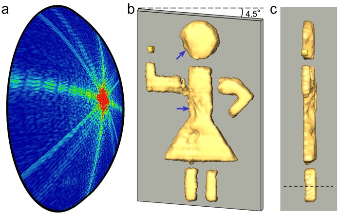
What the UCLA researchers realized was that the light recorded on the image detector was simultaneously seeing the object from many different directions. This process was actually a version of the stereoscopic vision that people have when they see with two eyes. The figure above is evidence of their success in determining the depth of an object. Panels b and c are 3D views of the stick girl from the front and side, respectively. Panel a is a picture of an Ewald sphere, a two-dimensional geometric construct used to show the relationship between the wavelengths of incident and diffracted X-ray beams as well as the diffraction angle for each reflection.
Miao and his colleagues showed that their new method not only works for the little stick girl, but also for computer-generated data simulating 3D reconstructions of a piece of sodium silicate glass and a single poliovirus. Kapteyn believes that with further development, ankylography could have applications in both the physical and life sciences, making it possible in the future to take 3D “snapshots” with nanometer resolution using an X-ray flash of only a few femtoseconds. Superman would be envious! - Julie Phillips